

Two researchers from the University Hospital and the Center for Applied Medical Research (CIMA) from the University of Navarra have published an article in Nature Cancer Reviews, one of the leading scientific journals in the area of cancer studies. The article, written by Ignacio Melero and Sandra Hervas-Stubbs, together with other scientists from the United States and Great Britain, addresses the use of a new pharmaceutical family with practical applications in cancer and chronic viral illnesses.

| Health & Medicine | March 6, 2007 10:01 PM |
For the first time in humans, scientists have found early indicators of inflammation — potential warning signs — in work-related injuries caused by repetitive motion.
| Full story | 0 Comments | 7712 views |

| Molecular & Cell Biology | March 6, 2007 09:01 PM |
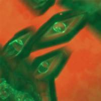
Ice crystals decorated by fluorescent antifreeze proteins. Antifreeze or “ice structuring” proteins – found in some fish, insects, plants, fungi and bacteria – attach to the surface of ice crystals to inhibit their growth and keep the host organism from freezing to death. Scientists have been puzzled, however, about why some ice structuring proteins, such as those found in the spruce budworm, are more active than others.
| Full story | 0 Comments | 5563 views |
| Molecular & Cell Biology | March 6, 2007 08:01 PM |
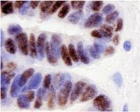
Ovarian cancer tissue showing PAX8 stained in black. A new study suggests that ovarian cancer cells form by hijacking a developmental genetic process normally used to form fallopian tubes. Scientists at the Georgia Institute of Technology and the Ovarian Cancer Institute discovered that the protein, PAX8, is involved in the development of fallopian tubes and is present in ovarian cancer cells, but not in normal ovarian tissue. The discovery not only provides a new target for diagnostic and therapeutic interventions, but also opens new avenues for basic research in ovarian cancer pathology. The research appears in Volume 104, Issue 3 of the journal Gynceologic Oncology.
| Full story | 0 Comments | 4454 views |

| Biotechnology | March 6, 2007 07:01 PM |
Leafy greens and beans aren't the only foods that pack a punch of folate, the vitamin essential for a healthy start to pregnancy.
| Full story | 0 Comments | 5567 views |

| Biology | March 6, 2007 06:26 PM |
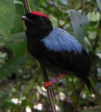
Adult male lance-tailed manakin on a branch Why do some individuals sacrifice their own self-interest to help others? The evolution and maintenance of cooperative behavior is a classic puzzle in evolutionary biology. In some animal societies, cooperation occurs in close-knit family groups and kin selection explains apparently selfless behavior. Not so for the lance-tailed manakin.
| Full story | 0 Comments | 5200 views |

| Environment | March 6, 2007 06:01 PM |
There's a scholarly debate brewing about whether pre-Columbian Amazonian populations settled in large numbers across Amazonia and created the modern forest setting that many conservationists take to be ‘natural.'
| Full story | 0 Comments | 5683 views |
| Molecular & Cell Biology | March 6, 2007 05:01 PM |
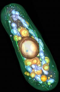
The electron tomogram of a complete yeast cell reveals the cellular architecture. It shows plasma membrane, microtubules and light vacuoles (green), nucleus, dark vacuoles and dark vesicles (gold), mitochondria and large dark vesicles (blue) and light vesicles (pink). Credit: Johanna Höög, EMBL Heidelberg Editor: A high resolution version of the picture is available!
Like our body every cell has a skeleton that provides it with a shape, confers rigidity and protects its fragile inner workings. The cytoskeleton is built of long protein filaments that assemble into networks whose overall architecture and fine detail can only be revealed with high resolution electron microscopy images. Researchers at the European Molecular Biology Laboratory (EMBL) and the University of Colorado have now obtained the first 3D visualization of a complete eukaryotic cell at a resolution high enough to resolve the cytoskeleton's precise architectural plan in fission yeast. The image of this unicellular organism will be published in this week's issue of the journal Developmental Cell and reveals remarkable insights into the fine structure of the cytoskeleton as well as its interactions with other parts of the cell.
| Full story | 0 Comments | 12427 views |

| Microbiology | March 6, 2007 04:26 PM |
UC Irvine researchers have combined genetic and geographic data of the H5N1 avian flu virus to reconstruct its history over the past decade. They found that multiple strains of the virus originated in the Chinese province of Guangdong, and they identified many of the migration routes through which the strains spread regionally and internationally.
| Full story | 0 Comments | 6167 views |
| Molecular & Cell Biology | March 6, 2007 02:26 PM |
Having an out-of-body experience may seem far-fetched to some, but for those with arousal system disturbances in their brains, it may not be a far off idea that they could sense they were really outside their own body watching themselves. In previous studies of more than 13,000 Europeans, almost 6 percent said they have had such an out-of-body experience.
| Full story | 0 Comments | 5716 views |
| Health & Medicine | March 6, 2007 12:26 PM |
Body mass index, or BMI, long considered the standard for measuring the amount of fat in a person's body, may not be as accurate as originally thought, according to new research.
| Full story | 0 Comments | 5007 views |
| Biotechnology | March 6, 2007 10:26 AM |
Using genetic manipulation to modify the activity of a plant enzyme, researchers at the U.S. Department of Energy's Brookhaven National Laboratory have converted an unsaturated oil in the seeds of a temperate plant to the more saturated kind usually found in tropical plants. The research will be published online by the Proceedings of the National Academy of Sciences (PNAS) the week of March 5, 2007.
| Full story | 0 Comments | 5041 views |


- In some genetic cases of microcephaly, stem cells fail to launch
- Yale team discovers how Zika virus causes fetal brain damage
- Scientists uncover the way a common cell enzyme alerts the body to invading bacteria
- Arctic gives clues on worst mass extinction of life
- Calcium channel blockers caught in the act at atomic level
Search Bio News Net
Free Biology Newsletter
- Flexibility That A.C.A. Lent to Work Force Is Threatened by...International Herald Tribune
- Bacteria Behind Legionnaires’ Disease Found at New York Police StationInternational Herald Tribune
- Prescription Price Crisis? 28 Million Americans See Spike in Drug...MSNBC.com: Health
- Seizing on Opioid Crisis, a Drug Maker Lobbies Hard for...International Herald Tribune
- Republicans are taking a big political risk on health careAP Health News
- Why We're So Divided Over Saving WolvesNational Geographic News