 Print Article
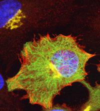
Parallel application of new (genetic tagging) and old targeting methods and fluorophores.An interdisciplinary team of biological imaging experts from the University of California, San Diego has published a review of fluorescent imaging technologies and underscored the importance of those technologies to major advances in the life sciences.
The article--"The Fluorescent Toolbox for Assessing Protein Location and Function"--is the cover story in the April 14 issue of the journal Science.
"Fluorescent imaging is critical to the observation of dynamic processes in living systems," said lead author Ben Giepmans, a research scientist in the UCSD-based National Center for Microscopy and Imaging Research (NCMIR). "Some of these techniques now also allow researchers to localize the responsible molecular machine in situ by electron microscopy."
Giepmans' co-authors on the Science paper include NCMIR director and UCSD School of Medicine neurosciences professor Mark Ellisman, pharmacology project scientist Stephen Adams, and Roger Tsien, professor of pharmacology, chemistry and biochemistry. The National Institutes of Health and the Howard Hughes Medical Institute supported work directly related to this review.
In their survey, the scientists contrasted the characteristic benefits and limitations of many new classes of fluorescent probes for studying proteins, including quantum dots, fluorescent proteins, and some genetic tags. Color-rich photomicrographs now routinely appear in scientific journals to illustrate dynamic biochemical processes. Those processes range from the expression of a specific gene to the redistribution of protein within a living cell.
Progress in developing new fluorescent probes over the last decade has been dramatic. "Whole new classes of fluorescent dyes, fluorescent proteins, and other hybrid probes are being engineered to illuminate specific biochemical structures and processes within living cells," said Ellisman. "They also make possible the direct correlated imaging of the underlying molecular complexes at higher resolution by electron microscopy."
Fluorescence imaging is rapidly becoming a biochemist's tool of choice for studying processes within living cells. Its rapid expansion is partially tied to a synergy of developments, including the increasing ease of implementing innovative targeting strategies to key cell metabolites and structures. Concomitant advances in instrumentation and data analysis are enabling scientists to identify and quantify dynamic biochemical processes of living cells under light and electron microscopes. Fluorescence techniques are being adapted for clinical and biochemical assays like biopsies and high-throughput drug screening, and are just beginning to find wider application in functional assays of living cells and animals.
Source : University of California - San Diego
 Mail to a Friend
