

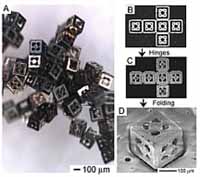
(A) Optical image showing a collection of biocontainers. (B-D) Optical and Scanning electron microscopy images at different stages of the fabrication process: (B) the 2D precursor with electrodeposited surfaces, (C) the precursor with surfaces and hinges, and (D) the self-assembled biocontainer. Johns Hopkins researchers have devised a self-assembling cube-shaped perforated container, no larger than a dust speck, that could serve as a delivery system for medications and cell therapy.

| Biotechnology | December 12, 2005 09:04 PM |
| Full story | 0 Comments | 5170 views |

| Environment | December 12, 2005 09:04 PM |

Loss of biodiversity in pollination networks may threaten the persistence of plant communities. (Photo: Fontaine et al.) As animal extinctions continue at the rate of one every 16 years, it's unclear how declining biodiversity will disturb ecosystem dynamics. Of special concern are the pollinators, essential players in the reproductive biology of plants, the earth's primary producers.
| Full story | 0 Comments | 7558 views |

| Microarray | December 12, 2005 09:04 PM |
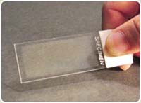
This slide includes approximately 2,000 combinations of eight enzymes used in human liver metabolism. To detect toxic drug compound reactions, the slide is “stamped” with a second slide of human organ cells. Photo by RPI/Moo-Yeal Lee and Jonathan DordickRecalls of popular prescription drugs are raising public concern about the general safety of new pharmaceuticals. A collaborative group of researchers says that identifying which drug candidates are toxic early in the discovery process can help prevent harmful pharmaceuticals from being placed on the market in the first place, and they have developed a tool to do it.
| Full story | 0 Comments | 6102 views |

| Biology | December 12, 2005 09:03 PM |
Safeguarding 595 sites around the world would help stave off an imminent global extinction crisis, according to new research published today in the Proceedings of the National Academy of Sciences (www.pnas.org).
| Full story | 0 Comments | 4262 views |

| Health & Medicine | December 12, 2005 09:03 PM |
For years, we have heard about the health benefits of taking low doses of aspirin – preventing everything from Alzheimer's disease to heart attacks and stroke. The news about aspirin just keeps getting better.
| Full story | 0 Comments | 5651 views |
| Biology | December 12, 2005 09:03 PM |
A study released today at the American College of Neuropsychopharmacology's Annual Meeting revealed that scientists have identified genes related to reaching age 90 with preserved cognition. The study, which was funded by the National Institutes of Health and conducted at the University of Pittsburgh, is among the first to identify genetic links to cognitive longevity.
| Full story | 0 Comments | 4203 views |

| Molecular & Cell Biology | December 12, 2005 09:03 PM |
A Yale School of Medicine study in the December issue of Lancet Oncology challenges mainstream oncology researchers to consider tumor cell hybridization with white blood cells as a major reason that cancer metastasizes or spreads to other parts of the body.
| Full story | 0 Comments | 3889 views |

- In some genetic cases of microcephaly, stem cells fail to launch
- Yale team discovers how Zika virus causes fetal brain damage
- Scientists uncover the way a common cell enzyme alerts the body to invading bacteria
- Arctic gives clues on worst mass extinction of life
- Calcium channel blockers caught in the act at atomic level
Search Bio News Net
Free Biology Newsletter
- Flexibility That A.C.A. Lent to Work Force Is Threatened by...International Herald Tribune
- Bacteria Behind Legionnaires’ Disease Found at New York Police StationInternational Herald Tribune
- Prescription Price Crisis? 28 Million Americans See Spike in Drug...MSNBC.com: Health
- Seizing on Opioid Crisis, a Drug Maker Lobbies Hard for...International Herald Tribune
- Republicans are taking a big political risk on health careAP Health News
- Why We're So Divided Over Saving WolvesNational Geographic News