 Print Article
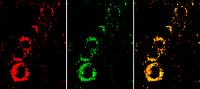
The yellow nanosensor signal in the overlay image (right) shows that the cells are active. If they were unhealthy, they would appear much redder. Center: the indicator dye signal. Left:... Countless mice, rats and rabbits die every year in the name of science – and the situation is getting worse. While German laboratories used some 2.41 million animals for scientific research in 2005, by 2009 this number had grown to 2.79 million. One third were destined for fundamental biology research, and the majority were used for researching diseases and developing medical compounds and devices. People demand medicines that are safe and therapies that are tolerable, but hardly anyone is happy to accept the need for animal testing. This is why scientists have spent years looking for methods that can replace them. Now researchers at the Fraunhofer Research Institution for Modular Solid State Technologies EMFT in Munich have found an alternative: they hope to use novel nanosensors to reduce the number of experiments that are carried out on animals. "We're basically using a test tube to study the effects of chemicals and their potential risks. What we do is take living cells, which were isolated from human and animal tissue and grown in cell cultures, and expose them to the substance under investigation," explains Dr. Jennifer Schmidt of the EMFT. If a given concentration of the substance is poisonous to the cell, it will die. This change in "well-being" can be rendered visible by the sensor nanoparticles developed by Dr. Schmidt and her team.
Cells – the tiniest living things – that are healthy store energy in the form of adenosine triphosphate (ATP). High levels of ATP are indicative of high levels of metabolic activity in cells. If a cell is severely damaged, it becomes less active, storing less energy and consequently producing less ATP. "Our nanosensors allow us to detect adenosine triphosphate and determine the state of health of cells. This makes it possible to assess the cell-damaging effects of medical compounds or chemicals," says Schmidt.
In order for the nanoparticles to register the ATP, researchers give them two fluorescent dyes: a green indicator dye that is sensitive to ATP, and a red reference dye that does not change color. Next, the scientists introduce the particles to living cells and observe them under a fluorescence microscope. The degree to which the particles light up depends on the quantity of ATP present. The more yellow is visible in the overlay image, the more active are the cells. If their health were impaired, the overlay image would appear much redder. "We could in future use cancer cells to test the effectiveness of newly developed chemotherapy agents. If the nanosensors detect a low concentration of ATP in the cells, we'll know that the new treatment is either inhibiting tumor cell growth or even killing them," says Schmidt. "The most promising agents could then be studied further."
The EMFT researchers' nanoparticles are extremely well suited to the task at hand: they are not poisonous to cells, they can easily pass through cell membranes, and they can even be directed to particular points where the effect of the test substance is of most interest. But before this procedure can be applied, it must first be approved by the regulatory authorities – so the EMFT experts have a long journey ahead of them to gain approvals from various official bodies. This prospect has not, however, stopped the researchers from refining the technology and coming up with new applications for it – for instance to test the quality of packaged meat and its fitness for consumption. To this end they have developed nanosensors that can determine concentrations of oxygen and toxic amines.
Source : Fraunhofer-Gesellschaft
 Mail to a Friend
