 Print Article
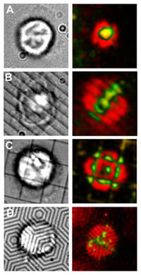
These fluorescently labeled electron micrographs show immunological synapses formed by T cell receptors (green) and adhesion molecules (red). Image (A) shows the synapse in its natural bull’s eye shape; in image (B) chromium lines were used to pattern the synapse with parallel lines; (C) the synapse was patterned into a square grid; and (D), the synapse was patterned into concentric hexagons.
An experiment that began as a "fantasy pipe dream" just three years ago is now a reality. Researchers with the Lawrence Berkeley National Laboratory (Berkeley Lab) and the University of California at Berkeley, combining nanotechnology with biochemistry, have created unique synthetic membranes that, for the first time ever, enable them to directly control signaling activity in living T cells from the immune system. Already their experiments have yielded surprising results.
"This marriage of inorganic nanotechnology with organic molecules and cells enables us to go inside a living cell and physically move around its signaling molecules with molecular precision," said Jay Groves, a chemist who holds a joint appointment with Berkeley Lab's Physical Biosciences Division and UC Berkeley's Chemistry Department. "Our experimental beaker has now become the inside of living cells and we can watch chemical reactions take place there."
Groves is the principal co-author, along with Michael Dustin, a cellular immunologist at New York University (NYU), of a paper published in the November 18, 2005 issue of the journal Science, entitled: "Altered TCR Signaling from Geometrically Repatterned Immunological Synapses." The lead author is Kaspar Mossman, a graduate student in Groves' research group, and the second co-author is Gabriele Campi, a graduate student at NYU with Dustin.
"Scientists, including ourselves, have been posing elaborate theories about how the strength and duration of signals that activate T cells are controlled by immunological synapses, without having been able to do direct experimentation of key factors," said Groves. "Three years ago, we had this fantasy pipe dream about an experiment to measure how alterations in the geometric shapes of the synapses – what we call spatial mutations – would affect T cell signaling. Then we realized, we have the tools to create nanoscale patterns, we can do this."
The human immune system is a remarkable collaboration of different types of cells, working together to protect our bodies from bacterial, parasitic, fungal or viral infections, and against the growth of tumors. The process starts when "antigens," special markers on the surface of a cell, identify another cell as "non-self," and signal the cellular warriors of the immune system to kill the invader. Leading this attack will be the T cells, lymphocytes from the thymus. It is well established that the key to T cell activation is the molecular signal coming off antigen-presenting cell surfaces. This signal must be enhanced and sustained long enough for the T cells to commit to mounting an immune response, and then must be cut off in time to avoid antigen-induced cell suicide or "apoptosis" of the T cells.
It has also been established that the control center for T cell signaling is at the junction or point of contact between T cells and antigens, dubbed the "immunological synapse" because it resembles the synapse between two communicating nerve cells. At the immunological synapse, a central cluster of T cell receptors surrounded by a ring of adhesion molecules form what co-author Dustin has described as a sort of "bull's-eye." The center of this bull's eye has been dubbed the "central supramolecular activation cluster," or c-SMAC, because it was believed to be the source of T cell activation.
"The original idea behind the c-SMAC was that the larger the T cell receptor cluster, the stronger the T cell activation signal," said Groves. "This simple vision of strength in numbers had begun to show cracks, and now we have demonstrated that just the opposite is true, the coalescence of the c-SMAC cluster extinguishes the T cell activation signal. The duration of the activation signal is related to the spatial organization of the T cell receptors rather than cluster size."
Groves and his colleagues constructed their synthetic membranes out of lipids which they assembled onto a substrate of solid silica so that the membranes were able to float freely a few nanometers above the substrate. This enabled the researchers to preserve the membranes in their naturally fluid state, allowing lipids and T cell receptor proteins to diffuse and interact freely over macroscopic distances.
"The fluidity of our membranes created artificial antigen-presenting cell surfaces that enabled the formation of functional immunological synapses with living T cells," said Groves.
Groves and his colleagues were able to spatially mutate the geometric shapes of the immunological synapses by embedding the silica substrate with chrome lines that were only 100 nanometers (about one ten-millionth of an inch) wide. These ultra-narrow chrome lines served as barriers that restricted the motion of membrane lipids and T cell receptor proteins. Using electron-beam lithography, the researchers were able to configure the chrome lines into several distinct patterns, including simple parallel lines, grids, and a series of concentric hexagons.
"By changing the shape of the immunological synapse, we showed that the synapse signal starts out in an amplified mode, and that the transport of the T cell receptors towards the center weakens and eventually extinguishes the signal, irrespective of the degree of clustering," Groves said. "This may help explain why diseases of the autoimmune system are so difficult to treat. T cell receptor proteins do not respond like a conventional target, where if you hit the bull's eye you trigger a signal. The spatial position of the receptor determines the type of signal it triggers."
If scientists can learn more about the impact that spatial arrangement has on the immunological synapse and its signaling strength, the information could benefit the future development of drugs for treating autoimmune diseases. Such information should also help scientists better understand the chemical language by which cells communicate with one another.
Groves said this new technique for spatial mutation studies should be applicable to many intercellular signaling systems. Already, he and his colleagues have begun applying it to study neuronal synapse formation, and cell signaling mechanisms in the development of cancer. They are also using it to look at the dynamic range of signaling over which T cell receptors can respond.
"Essentially, these experiments amount to using inorganic nanotechnology to physically grab a protein in a living cell and move it to another position in that cell – then watch how the cell responds," said Groves. "We used it to study the T cell as a paradigm system, but the theme here is much more general. Whereas the spatial position of molecules is rarely thought to play an important role in the outcome of a chemical reaction, with our experimental technique we are seeing that, in living cells, this is not the case. The spatial position encodes information which can be directly translated into altered chemical outcomes."
The earliest indications that spatial positions could influence T cell signaling and that the synaptic pattern might actually help to extinguish the signal came from the work of Arup Chakraborty, a chemical engineering professor who, at the time, held a joint Berkeley Lab/UC Berkeley appointment and is now with MIT University. Chakraborty is a pioneer in the use of computer simulations, called "experiments in silico," for studying important problems in cellular immunology. In 2003, his computational models indicated that the immunological synapse is responsible for intense but self-limited T cell signaling.
Source : DOE/Lawrence Berkeley National Laboratory
 Mail to a Friend
