 Print Article
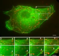
These time-lapse images of a bovine aortic endothelial cell reveal the motion toward the cell's nucleus of a message-carrying protein called paxillin (orange) in tandem with actin filaments (green). Credit: UC San Diego Scientists have captured on video the intracellular version of a postal delivery service. Reporting in the journal Biochemical and Biophysical Research Communications (BBRC), bioengineering researchers at UC San Diego published videos of a key message-carrying protein called paxillin moving abruptly from hubs of communication and transportation activity on the cell surface toward the nucleus. Paxillin was labeled with a red fluorescence marker to make it stand out in live cells.
Video available at http://video-jsoe.ucsd.edu/asx/Paxillin.paper.wmv.asx
“It’s amazing to us. We thought the cell was so simple,” said Shu Chien, the senior author of the BBRC paper and a professor of bioengineering at UCSD’s Jacobs School of Engineering. “But it’s really very complex and I’m not sure we’re covering much as yet. We certainly don’t know all the interactions among these molecules that bring the cell into action.”
Examining living cells through a microscope, Chien and the paper’s co-author, associate project scientist Ying-Li Hu, filmed red-fluorescence-tagged paxillin molecules traveling from cells’ outer membrane along green-fluorescence-labeled traces of cytoskeleton. Even without video evidence, scientists have confirmed over the past 10 years that higher organisms use paxillin as a transmitter of locomotion and gene-expression signals from several classes of growth-factor receptors to the nucleus.
Cancer researchers are eager to understand paxillin’s many interactions because their malfunctions have been linked to a variety of cancers, tumor metastasis, and other disease processes. Tumor-causing versions of signaling molecules may attach to paxillin and disturb the normal adhesion and growth factor signaling steps required for controlled proliferation and cell growth. For example, human papilloma virus, which can cause cervical cancer, makes a protein that binds to paxillin and that interaction may contribute to the carcinogenic potential of the sexually transmitted virus.
Chien and Hu obtained cells for their most recent study from the inner membranous lining of a cow aorta. They added genetically engineered proteins tagged with red and green fluorescence markers to the bovine aortic endothelial cells, grew them in tissue culture, and filmed them while they passed fluid across the cells’ surface in a simulation of flowing blood. With that physical stimulus, paxillin consistently moved in the same direction as the fluid flow.
Paxillin is found primarily at focal adhesions, busy intersections of activity scattered around the cell’s cytoplasmic membrane. Focal adhesions are regions rich with receptors for growth factors and they are also structural attachment points that link the extracellular world to the protein filaments and tubes that comprise a cell’s internal cytoskeleton.
While researchers showed that paxillin serves as a docking station for a variety of signaling and structural proteins, they have been limited to visualizing paxillin with dyes that stain cells rigidly attached to microscope slides. Those snapshots of fixed cells can’t reveal paxillin movement.
“We can now see the dynamics of how paxillin and other proteins move inside the cell with new fluorescent labels and live-cell video microscopy,” said Chien, director of the Whitaker Institute of Biomedical Engineering at the Jacobs School.
The two-color labeling technique of Chien and Hu revealed that red-labeled paxillin is linked to green-labeled actin filaments, the thinnest of three types of protein filaments that make up the cytoskeleton of cells of higher organisms. Paxillin either slides along the actin filaments or is pulled by them. In a surprise, Chien and Hu also showed that paxillin itself forms long, fibrous structures in the cell cytoplasm.When actin is chemically removed, paxillin no longer moves and its fibrous structures disappear.
“As least by showing that paxillin and actin filaments move together we have some insight into the possibility of how paxillin can transmit information from one region of the cell, such as the periphery, to another region of the cell, such as the nucleus,” said Chien. “The whole situation is much more complex than we have been able to show.”
Chien, Hu, and their colleagues at UCSD had previously reported that paxillin can be rapidly assembled and disassembled, and they hypothesize that this process enables cell migration by assembling new focal adhesion sites in the direction of cell movement and disassembling them on the trailing side of the cell. Researchers are applying the fluorescence-labeling technique to the many other signaling and structural proteins found predominantly at focal adhesions.
“When we piece all of these things together we can get a more complete understanding of how the cell functions, both in migration and signal transduction,” said Chien. “If we can understand the details of these processes, we can not only understand how normal cells function, but we can also look at diseased cells to see why they sometimes don’t move properly or why they don’t transmit information properly.”
Source : University of California - San Diego
 Mail to a Friend
