 Print Article
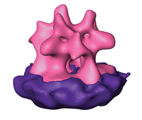
3-D structure of the protein complex that allows the retrovirus to penetrate living cells. Retroviruses are among the trickier and more malicious disease agents, causing AIDS and cancers such as leukemia. The viruses manage to sneak into cells with the help of special protein assemblies scattered all over their surfaces. These retrovirus surface proteins cause the membrane envelope of the virus to fuse with the membrane of the cell, spilling virus RNA into the cell to wreak damage. Now, a team of scientists at the Weizmann Institute of Science and the Max Planck Institute for Biochemistry has obtained a close-up 3-D portrait of the large protein complex on the virus that enables its entry into the cell. Their work appeared in the Proceedings of the National Academy of Sciences, USA in March.
These protein complexes recognize and bind to specific sites on the cellular membrane and mediate the fusion process, the first steps in virus infection. However, the shape of this complex on retroviruses and the way that it works had long evaded efforts at detection by various scientific groups. The difficulty is that crystallization, the leading method of preparing proteins for structure solving, does not work well with the elaborate, envelope-bound complexes, which tend to fall apart when they are removed from the virus membrane. Dr. Deborah Fass of the Weizmann Institute's Structural Biology Department had managed to determine the structures of assorted parts of the complex in the past, but needed a better understanding of how the complex works as a whole.
To accomplish her goal, Fass and student Nathan Zauberman teamed up with scientists from Max Planck's Molecular Structural Biology Department in Martinsried, Germany to try an alternative method of getting an image of the complex. They turned to the electron microscope, a standard tool for observing larger structures such as cell sections.
Viewing a single, relatively small protein complex was pushing the limits of this technology, but the Max Planck group, expert at developing both the hardware and the software required for visualizing biological structures using electron microscopy, was up to the task. The technique used, known as cryo-electron tomography, involved quick-freezing the viruses in liquid ethane, capturing snapshots of them at various angles and then combining the snapshots to create three-dimensional pictures. From dozens of these digitized 3-D pictures of whole viruses, hundreds of protruding surface protein complexes could be cut out, aligned, and averaged. Though the resulting image did not have quite as high a resolution as those obtained through crystallography, it allowed the scientists to get a complete and fairly detailed picture of this important protein complex in its natural environment. "After years of trying to imagine how the pieces fit together, suddenly we had the real structure right in front of us. Some aspects of it looked familiar, but others were completely unanticipated," says Fass.
The scientists were surprised to note that the shape of the complexes on the retroviruses bore little resemblance to other known viral envelope protein structures such as those on flu viruses. They also saw strong evidence that the protein complex undergoes a radical change in shape and arrangement of its component parts as it attaches to cells and initiates membrane fusion. Fass was able to see how a smaller protein piece she had previously isolated and analyzed by crystallization fit into the whole, giving her further clues as to how the virus locks onto the cell membrane.
The retrovirus used by Fass and the team is similar to that which causes leukemia in humans. They hope, with further research, to understand the conformational changes the envelope protein complex undergoes as it works, and to find ways to stop those changes from taking place.
Dr. Debora Fass' research is supported by the Clore Center for Biological Physics; the Helen and Milton A. Kimmelman Center for Biomolecular Structure and Assembly; and the Leukemia Research Foundation.
Dr. Fass is the incumbent of The Lillian & George Lyttle Career Development Chair.
Source & Image : Weizmann Institute
 Mail to a Friend
